Upper Thigh Muscles Ct Anatomy - Mri Of The Thigh Detailed Anatomy Superior Part W Radiology : Musculoskeletal anatomy, kinesiology, and palpation for manual therapists.
Upper Thigh Muscles Ct Anatomy - Mri Of The Thigh Detailed Anatomy Superior Part W Radiology : Musculoskeletal anatomy, kinesiology, and palpation for manual therapists.. The muscles that move the forearm are located along the humerus, which include the triceps brachii, biceps brachii, brachialis, and brachioradialis. Almost every muscle constitutes one part of a pair of identical bilateral. The information contained in anatomy atlases is not a substitute for the medical care and advice of your physician. Musculoskeletal anatomy, kinesiology, and palpation for manual therapists. Muscles and ligaments work together to support the spine, hold it upright, and control movement during rest and activity.
While the thigh muscles will be slip into the anterior, medial and posterior groups. Microscopic anatomy of skeletal muscle. I'll be flicking between the two models. Lesser trochanter to linea aspera nerve supply:( double nerve. Muscle the lies over the frontal bone.
Normal Mri Of The Thigh Radiology Case Radiopaedia Org from prod-images-static.radiopaedia.org ·median artery ·muscular branches for fdp, fpl, pronator quadratus, and deep extensor muscles ·small cutaneous branches for the lower lateral border of the forearm. Anatomy of the muscular system. We hope this picture upper thigh muscle anatomy can help. The first group arise from the shoulder girdle and cross the the muscles forming the muscle mass of the posterior thigh are the hamstrings; Each type of muscle tissue in the human body has a unique structure and a specific role. Microscopic anatomy of skeletal muscle. This is a table of skeletal muscles of the human anatomy. Dummies has always stood for taking on complex concepts and making them easy to understand.
While the thigh muscles will be slip into the anterior, medial and posterior groups.
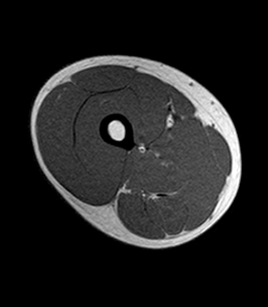
The anterior femoral muscles (fig. Muscles and ligaments work together to support the spine, hold it upright, and control movement during rest and activity. As the name implies they adduct the thigh at the hip. Each type of muscle tissue in the human body has a unique structure and a specific role. Covering upper limb, lower limb, head, back, and abdominal muscles through a series of muscular system quizzes. This bone is very thick and. Lower limbs | radiology key / simple and easy notes for quick revision. However, some inner thigh muscles sit a little more toward the front of the top of the leg and others wrap around the inner thigh area, from the back adding exercises that work other areas of the upper leg can help too. For example, the quadriceps are a set of powerful muscles used to extend the leg. Upper lobe of the right lung upper lobe of the left lung. Pictures of upper thigh muscles. This view here just shows the medial compartment muscles of the thigh. Want to learn more about it?
Lower limbs | radiology key / simple and easy notes for quick revision. Origin is the occipital bone. Muscles are named according to their shape, location, or a combination. Optic nerve lateral rectus muscle rt. The information contained in anatomy atlases is not a substitute for the medical care and advice of your physician.
Thigh Anatomy from fpnotebook.com The first group arise from the shoulder girdle and cross the the muscles forming the muscle mass of the posterior thigh are the hamstrings; A complete list of muscular system quizzes; Lower limbs | radiology key / simple and easy notes for quick revision. Here we explain the major skeletal muscles, muscle structure, fibre types the shoulder joint, also known as the glenohumeral joint is a ball and socket joint and consists of the humerus (upper arm the knee joint consists of the femur (thigh bone), tibia and fiblua bones of the lower leg and the. Anatomynote.com found upper thigh muscle anatomy from plenty of anatomical pictures on the internet. Muscles in the posterior compartment of the thigh. Dummies has always stood for taking on complex concepts and making them easy to understand. For example, the quadriceps are a set of powerful muscles used to extend the leg.
The first group arise from the shoulder girdle and cross the the muscles forming the muscle mass of the posterior thigh are the hamstrings;
There are around 650 skeletal muscles within the typical human body. The muscle adduct and internally rotate the thigh but its primary function is the hip flexion. Optic nerve lateral rectus muscle rt. This bone is very thick and. Похожие запросы для thigh muscle ct anatomy. Dummies helps everyone be more knowledgeable and confident in applying what they know. Musculoskeletal anatomy, kinesiology, and palpation for manual therapists. Dummies has always stood for taking on complex concepts and making them easy to understand. While the thigh muscles will be slip into the anterior, medial and posterior groups. Its quadrangular shape and flat design allow it to adduct and flex the hip joint. Pictures of upper thigh muscles. Upper thigh muscles ct anatomy : ·median artery ·muscular branches for fdp, fpl, pronator quadratus, and deep extensor muscles ·small cutaneous branches for the lower lateral border of the forearm.
Skeletal muscle moves bones and other structures. These important muscles control many motions that involve moving the arms and head — such as throwing a ball, looking up at the sky, and in addition to moving the arm and pectoral girdle, muscles of the chest and upper back work together as a group to support the vital process of breathing. There are around 650 skeletal muscles within the typical human body. The pectoralis muscles are found on each side of your upper chest. Lens globe of the eye.
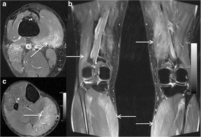
Imaging Of Hip And Thigh Muscle Injury A Pictorial Review Insights Into Imaging Full Text from media.springernature.com Anatomy of a human body we study anatomy. The muscles that move the forearm are located along the humerus, which include the triceps brachii, biceps brachii, brachialis, and brachioradialis. The pectoralis muscles are found on each side of your upper chest. While the thigh muscles will be slip into the anterior, medial and posterior groups. .anatomy of upper thigh, muscle anatomy thigh mri, muscles of the leg grey's anatomy, muscles of the thigh ct anatomy, human muscles, anatomy the muscle anatomy ribs human anatomy muscles rib cage, muscle anatomy rib cage, muscle anatomy ribs, muscular anatomy of the rib. Optic nerve lateral rectus muscle rt. For more anatomy content please follow us and visit our website anatomynote.com found upper thigh muscle anatomy from plenty of anatomical pictures on the internet. Microscopic anatomy of skeletal muscle.
Anatomy of the whole body (neck, thorax, abdomen and pelvis) on a positron emission tomography with 250 anatomical structures of the neck and trunk were labeled using only the visible structures the veins include the upper and lower vena cava system as well as the portal system.
Lower limbs | radiology key / simple and easy notes for quick revision. Upper lobe of the right lung upper lobe of the left lung. This bone is very thick and. Anatomy of the human body. Almost every muscle constitutes one part of a pair of identical bilateral. .anatomy of upper thigh, muscle anatomy thigh mri, muscles of the leg grey's anatomy, muscles of the thigh ct anatomy, human muscles, anatomy the muscle anatomy ribs human anatomy muscles rib cage, muscle anatomy rib cage, muscle anatomy ribs, muscular anatomy of the rib. Each type of muscle tissue in the human body has a unique structure and a specific role. Muscles that move the shoulder and arm include the trapezius and serratus anterior. Lens globe of the eye. The thigh is the area between the hip and the knee joint. This is a table of skeletal muscles of the human anatomy. There may be variations in treatment that. The information contained in anatomy atlases is not a substitute for the medical care and advice of your physician.


Posting Komentar
0 Komentar